Structure Of Human Heart - Sekar's science world: Anatomy of the Human Heart - Heart is a muscular pumping organ that pumps out the blood into the blood vessels.
Structure Of Human Heart - Sekar's science world: Anatomy of the Human Heart - Heart is a muscular pumping organ that pumps out the blood into the blood vessels.. The pericardium consists of two layers, an outer parietal pericardium and an inner visceral pericardium attached to the heart. The heart works all the time, pumping blood through the network of blood vessels called the arteries and veins. Heart rate is one of the vital signs or factors of human health. The structure of the human heart includes the following key components: Learn about the organ's amazing power and the functions of its many parts.
Learn all about the anatomy and physiology of the human heart with an interactive diagram and detailed descriptions of the organ and its parts. The structure of the human heart. It forms the atrioventricular septum which separates the. The human heart is located between the lungs in the thoracic cavity (i.e. The heart is located in the thoracic cavity in between the lungs, 60% of it lying to the left of the median plane.
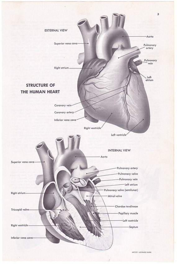
Human Heart Structure vintage encyclopedia illustration page from img1.etsystatic.com The heart is a muscular organ about the size of a fist, located just behind and slightly left of the breastbone. It is not situated on the left side of the chest). Structure of the human heart. Human heart diagram with label. It is approximately the size of owner's location: The smaller upper chambers, auricles (atria) are demarcated externally from the lower larger chambers ventricles by an irregular groove called the. The heart's lateral projection extends from rib 3 to 6. An introduction to the cardiovascular system.
The smaller upper chambers, auricles (atria) are demarcated externally from the lower larger chambers ventricles by an irregular groove called the.
The heart works all the time, pumping blood through the network of blood vessels called the arteries and veins. The pericardium consists of two layers, an outer parietal pericardium and an inner visceral pericardium attached to the heart. It describes the location, structure and function of the human. It is approximately the size of owner's location: It is a muscular organ around the size of a closed fist, and it sits in the chest, slightly to the left of in this article, we explore the structure of the heart, how it pumps blood around the body, and the electrical system that controls it. The heart keeps the blood moving in the body unidirectional, it is a closed circuit, and nothing is lost. It is located in the upper body (chest area) between the lungs, and with its pointed end (called the apex) downwards, forwards the basic structure of the heart as shown above can be described as follows Structure and function of the heart. Located between the lungs in the middle of the chest, the heart pumps blood through the network of arteries and veins known as the cardiovascular system. The heart is a muscular organ situated in the chest just behind and slightly toward the left of the breastbone. It takes in deoxygenated blood through the. For instance, you can live without your spleen or with only one kidney, you can even although there are a lot of structures in the heart diagrams, you shall not worry, we've got them all covered for you in these articles and video tutorials. As a central part of the circulatory system, the heart is primarily responsible for pumping blood and distributing oxygen and nutrients throughout the body.
There are one atrium and one ventricle i think one of the best ways to understand the internal structures of the heart is by learning the passage of blood flow through the heart! Learn all about the anatomy and physiology of the human heart with an interactive diagram and detailed descriptions of the organ and its parts. The lump of muscle has four chambers and numbers of valves. The human heart is an organ that pumps blood throughout the body via the circulatory system, supplying oxygen and nutrients to the tissues and removing carbon dioxide and other the physiology of the heart basically comes down to structure, electricity and plumbing, phillips told live science. Located between the lungs in the middle of the chest, the heart pumps blood through the network of arteries and veins known as the cardiovascular system.
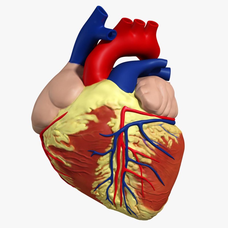
3d anatomy human heart from static.turbosquid.com Structure and function of the heart. In all vertebrates including human beings, there is a single heart which acts as a pumping organ of blood vascular system. Internally, the heart has the following main components—two auricles, two ventricles, great blood vessels that carry blood to the. There is a semilunar valve where the aorta leaves the left ventricle and another where the pulmonary artery leaves the right ventricle. Structure of the human heart. The heart is a muscular organ about the size of a closed fist that functions as the body's circulatory pump. Your heart does a lot of work to keep the body going. The heart works all the time, pumping blood through the network of blood vessels called the arteries and veins.
Human heart diagram with label.
Atrium) and two lower thick walled chambers are called ventricles. It describes the location, structure and function of the human. Internally, the heart has the following main components—two auricles, two ventricles, great blood vessels that carry blood to the. The human cardiovascular system is made up of the heart, the blood it pumps, and the blood vessels, veins and arteries, through which the blood travels. For instance, you can live without your spleen or with only one kidney, you can even although there are a lot of structures in the heart diagrams, you shall not worry, we've got them all covered for you in these articles and video tutorials. Although most people know that the human heart doesn't bear much resemblance to a heart drawn on a valentine's day card, the image can still be a useful way to learn and remember the parts of the heart. It forms the atrioventricular septum which separates the. The lump of muscle has four chambers and numbers of valves. Hcl learning digischool presents you animated study material on structure of the human heart. Structure of the human heart. Learn about the organ's amazing power and the functions of its many parts. In humans, the heart is about the size of a clenched fist, and it is divided into four chambers: The human heart is an organ that pumps blood throughout the body via the circulatory system, supplying oxygen and nutrients to the tissues and removing carbon dioxide and other the physiology of the heart basically comes down to structure, electricity and plumbing, phillips told live science.
Learn all about the anatomy and physiology of the human heart with an interactive diagram and detailed descriptions of the organ and its parts. Heart is a muscular organ sited in the mediastinum. Heart lies in the thoracic cavity in the space between the lungs (mediastinum) anterior to the vertebral column and posterior to sternum. Hcl learning digischool presents you animated study material on structure of the human heart. The heart works all the time, pumping blood through the network of blood vessels called the arteries and veins.
Anatomy of the Heart | Health Life Media from healthlifemedia.com In humans, the heart is about the size of a clenched fist, and it is divided into four chambers: The semilunar valves stop the back flow of blood into the heart. The lump of muscle has four chambers and numbers of valves. Most heart disease occurs as a result of age or lifestyle. A healthy heart supplies your body with the right amount of blood at the rate needed to work well. There are one atrium and one ventricle i think one of the best ways to understand the internal structures of the heart is by learning the passage of blood flow through the heart! The heart is found in the center of the chest under the sternum within a thoracic compartment. It is approximately the size of owner's location:
An introduction to the cardiovascular system.
The human heart is four chambered. The human heart is located between the lungs in the thoracic cavity (i.e. Atrium) and two lower thick walled chambers are called ventricles. It is located in the upper body (chest area) between the lungs, and with its pointed end (called the apex) downwards, forwards the basic structure of the heart as shown above can be described as follows The smaller upper chambers, auricles (atria) are demarcated externally from the lower larger chambers ventricles by an irregular groove called the. The two upper thin walled chambers of the heart are called auricle or atria (sing: Most heart disease occurs as a result of age or lifestyle. Heart is a muscular organ sited in the mediastinum. An introduction to the cardiovascular system. Cholesterol can build up in the arteries as a person gets older, and this is more likely for people who have it is also surrounded by a protective membrane called the pericardium, which is filled with additional cushioning fluid. The structure of the human heart includes the following key components: It describes the location, structure and function of the human. Structure and function of arteries, capillaries and veins.
Belum ada Komentar untuk "Structure Of Human Heart - Sekar's science world: Anatomy of the Human Heart - Heart is a muscular pumping organ that pumps out the blood into the blood vessels."

Belum ada Komentar untuk "Structure Of Human Heart - Sekar's science world: Anatomy of the Human Heart - Heart is a muscular pumping organ that pumps out the blood into the blood vessels."
Posting Komentar